info@jtmopticians.co.uk
0191 4561552


The Advanced Eye Examination involves three main components: OCT scan, Retinal Photography and Anterior eye Digital Slit Lamp Imaging.
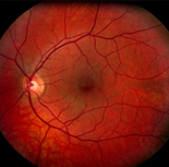
involves taking a photograph of the back of the eye so that we can get a detailed assessment of the Optic Nerve Head, Blood vessels & Macula. This is important for detecting & monitoring retinal eye conditions & general health conditions.
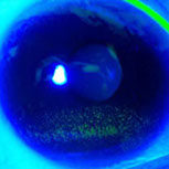
can assess the front of the eye including the eyelids, tear film, corneas & lens. This can be really useful for monitoring & treating common conditions such as blepharitis (inflammation for the eyelids) & dry eye. We can also monitor cataract development in the lens and show you how it changes from year to year.

Our REVO FC OCT scanner gives an extremely fast, non-invasive and accurate assessment of deep retinal layers. You could think of it as the MRI for the eye.


